Cryptophasa lasiocosma (Lower, 1908)
+Kurandra+Qld+F.jpg)
♂ - Qld, 1 mile N of Kuranda 1200 ft Alt., 23. Apr. 1969, I.F.B. Common M.S. Upton leg. (ANIC). [AMO].
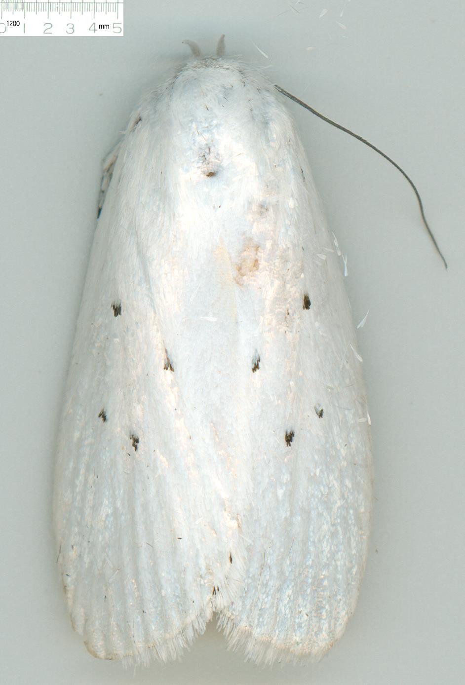
♀ - gvc6170, Herveys Range, Queensland. Collected by Graeme Cocks.

♀ - gvc6170, Herveys Range, Queensland. Collected by Graeme Cocks.

♀ - gvc2949, Herveys Range, Queensland. Collected by Graeme Cocks.
Cryptophaga lasiocosma Lower, 1908. New Australian Lepidoptera. No. XXV. Transactions of the Royal Society of South Australia 32: 110–120 [117]. Holotype SAMA ♀, Kuranda, Qld.
Cryptophasa pentasticta Turner, 1917. Lepidopterological gleanings. Proc. R. Soc. Qd 29: 70–106 [91]. Holotype ANIC ♀, Kuranda, Qld.
Cryptophasa lasiocosma Lower. Birket-Smith, 1974, Morphology of the male genitalia of Lepidoptera. III Appendix Cryptophasa lasiocosma (Xyloryctidae). Ent. scand. 5 184-188.
Cryptophasa lasiocosma Lower. Kanazawa, Itaru, 1987, The male genital morphology of Metathrinca tsugensis (Kearfott) with special remarks on the formation of the "valvella", Bulletin of the Osaka Museum of Natural History, 41 (7-15).
Cryptophasa lasiocosma (Lower 1908) [ = pentasticta, syn. n.]. Common, in Nielsen, Edwards, & Rangsi, 1996, Checklist of the Lepidoptera of Australia. Monographs on Australian Lepidoptera, 4: i-xiv, 1-529 & CD-ROM [86].
Cryptophasa lasiocosma Lower. Kristensen, ed., 2003, Handbook of Zoology, Volume IV, Arthropoda: Insecta, Part 36, Lepidoptera , Moths and Butterflies Vol, 2, Morphology, Physiology, and Development, pp. 101. 105, 110.
Cryptophasa lasiocosma (Lower 1908). Beccaloni, G. W., Scoble, M. J., Robinson, G. S. & Pitkin, B. (Editors). 2003. The Global Lepidoptera Names Index (LepIndex). World Wide Web electronic publication. http://www.nhm.ac.uk/entomology/lepindex [accessed 16 April 2010].
Cryptophasa pentasticta Turner, 1917. Beccaloni, G. W., Scoble, M. J., Robinson, G. S. & Pitkin, B. (Editors). 2003. The Global Lepidoptera Names Index (LepIndex). World Wide Web electronic publication. http://www.nhm.ac.uk/entomology/lepindex [accessed 16 April 2010]. [Synonymy not noted].
Cryptophasa lasiocosma (Lower 1908). Edwards, E. D. (2003), Xyloryctinae. Australian Faunal Directory. Australian Biological Resources Study, Canberra. http://www.environment.gov.au/biodiversity/abrs/online-resources/fauna/afd/taxa/XYLORYCTINAE [accessed 18 June 2010].
Original description, Lower, 1908
Cryptophaga lasiocosma, n. sp.
♀, 54 mm. Head, palpi, antennae, thorax, legs, and abdomen snow-white, antennae fuscous on terminal 2/3, thorax ochreous anteriorly, abdomen with reddish segmental margins, first segment broadly banded with orange ochreous. Tarsi banded with black. Forewings elongate, moderate, costa gently arched, termen rounded, oblique, 2 from 2/3, shining snow white; markings black, a spot near base; a larger one in cell at 2/5, and another on fold in middle; one or two black scales near tornus; cilia shining snow white. Hindwings and cilia shining snow white.
Kuranda, Queensland. One specimen, from Mr. F. P. Dodd; bred in December.
Nearest Argyrias, Turn., but differs in the discal dots, clear white hind wings, etc.
Synonomic description, Turner 1917
Cryptophasa pentasticta, n. sp.
πενταστιχτος, five-spotted.
♀ 43 mm. Head, thorax, and abdomen white. Palpi moderate, second joint reaching base of antennae, terminal joint 2/5; white, basal half of external surface of second joint black. Legs white; tarsi broadly annulated with black; inner surface of anterior femora and tibiae black. Forewing moderate, costa slightly arched, apex obtuse, termen slightly oblique, rounded beneath; 2 from ¾; white; five black dots, near base, second at 2/5, third on fold beyond middle, fourth at 3/5, fifth beyond and beneath fourth; two or three blackish terminal dots above tornus; cilia white. Hindwings and cilia white.
N.Q., Kuranda near Cairns in July; one specimen taken by Miss Enid Hewett, of Melbourne, communicated to me by Mr. F. P. Dodd.
Other references
Abstract
The genitalia or a male of the Australian moth Cryptophasa lasiocosma Lower have been dissected. The valves and particularly the phallic apparatus showed some morphologically very primitive traits, which place it close to the hitherto hypothetical intermediate form in respect of the formation of an aedoeagus from the valvellae.
In a previous paper (Birket-Smith, 1974a) the writer figured a hypothetical, intermediate stage in the formation of the aedoeagus from the derivatives of the mesapophyses, the valvellae, suggesting that these together formed a supporting tube enclosing a primitive, intromittant organ, the penis (1.c fig. 14 B). In the same paper the genus Aeolanthes Meyrick (Xyloryctidae) was mentioned as an example of a form in which the valvellae were forming a support for phallos, as far as could be judged from figures by Clarke (1955). Meantime the acquisition of a fresh specimen of Cryptophasa lasiocosma Lower, another member of the Xyloryctidae, has made an examination of the musculature of the male genitalia of a species of this family possible. These genitalia turned out to be of great morphological interest, particularly the phallos which is very similar to the above mentioned, hypothetical form. A short description of the anatomy and morphology of these genitalia will therefore be given below.
The specimen was caught on mercury vapour light in rain forest by Cairns, Queensland on 2 Sept. 1972, dried, and stored in paradichloro benzene. In the laboratory the genitalia were removed and softened in phenol alcohol (Birket-Smith, 1959, 1965) .
Superficially the genitalia look quite ordinary, maybe except for the uncus.
The tegumen, te, is a rather broad, triangularish crescent. Caudally it is by a flexible zone connected to the anal complex that consist of a dorsal uncus, uc, and a ventral subscaphium (“gnati”?), ss, between which the hardly protruding anus, as, is situated. The uncus, uc, is double with a straight, wedge-shaped process, and a ventral, stout hook with a bifid tip. The uncus, uc, is moved by a single. fiat, and broad muscle, m.1, originating on the antecosta of the tegumen, te, and inserted on the ventral edge of uncus, uc, just above the anus, as.
The subscaphium (“gnati”'?), ss, is V-shaped and synsclerituous with uncus, the ‘arms’ of the V connected laterally to the base of uncus through an intricate system of folds, that allows easy mutual movements; ventrally the subscaphium is drawn out into a laterally compressed, curved process. A pair of long muscles, m.2, originate laterally on tegumen, te, caudally of its antecosta, and are inserted laterally on the subscaphium, ss; frontally the fibers of these muscles lie close against those of m.1. The gnati are defined as derivatives of the tenth sternum (Klots, 1970: 258). As far as the present subscaphium is concerned, no indication of it being a derivative of the tenth sternum has been found. On the contrary, both its synsclerituous connection to, and the origin of its muscles on the tegumen-uncus complex - which is derived from the ninth primary segment - speak against this conception. The term subscaphium indicating a secondary sclerotization has thus been preferred here.
The vinculum, vi, is formed from a pair of slender, lateral. nearly horizontal arms connected to a broad, and double, ventral piece, the saccus, sa.
The valve is relatively long and slender. On the middle of its ventral edge is a distinct notch from which a faint line runs dorsad on the lateral side of the valve, separating the more strongly sclerotized basal part, bv, from the softer, distal part, sv. On the medial side of the valve the notch continues as the distal delimitation of the - on this side swollen -basal half of its ventral edge (“sacculus” ). From the distal end of this swelling protrudes dorsally a strongly sclerotized, dorsad curved process (“clasper”), av, which is slightly concave on its dorsal side, particularly so at its base; the medial edge of this concavity continues as a fold to base of the valve. From this fold to the ventral edge of the valve including the swelling on the medial side of it the valve is thinly sclerotized, while the remainder of its medial surface is membranous. Proximally the process, av, is relatively thick and the lateral side of its base is nearly touching the lateral side of the valve.
Three extrinsic muscles are connected to the valve, viz. m.3 originating laterally on tegumen, te, and inserted dorsally on the proximal edge of the valve, and m.4 originating laterally on vinculum and inserted just ventrally of m.3. Further the phallic protractor, m.6, which originates laterally on the phallos internus, pi, and is inserted mainly on the lateral wall of the basal part of the valve, bv, but a few of the dorsalmost fibres are inserted on the lateral side of the base of the curved process, av. The extrinsic position of m.6 is a secondary one, primitively it is an intrinsic muscle of the valve (Birket-Smith. 1974b). Of obviously intrinsic muscles the valve contains only one, viz. m.5 originating on the lateral side of the basal part of the valve, bv, and inserted on the medial side of the base of the curved process, av.
Referring to an earlier paper on the subject (Birket-Smith, 1974b) the basal part, bv, of the valve is considered the basis valvae, a derivative of the gonocoxa, and the curved process, av, the ala valvae, a derivative of the gonostylus. The musculature of ala valvae must be considered a very primitive trait, since m.5 is well developed and has retained both of its primitive insertions and m.6 has still retained some of its primitive insertion on the ala valvae, even if most of it now has acquired a secondary insertion on the basis valvae. Also the large basis valvae must be considered a primitive trait. The remainder, soft part of the valve, sv, is the supra valva, a mere outgrowth on basis valvae.
The phallos is the most complicated but also the most interesting structure. The ductus ejaculatorius, de, in its center continues distally as an eversible vesica, ve. Distally the membrane of the vesica is everted; it passes imperceptibly into the external wall of the penis, pn, a weakly sclerotized tube. that proximally is continuous with the internal wall of the distinctly double-walled phallos interna, pi. In the proximal fourth of the penis, the two walls of the phallos interna are separated by a narrow, air-filled space, the subgenital crypt, cr; distally of this the two walls are fused for a short distance, possible indicating the primitive zona. A pair of latero-ventral muscles, m.15, originate in phallos interna, pi , and are inserted proximally on vesica, ve, acting as a retractor of the latter.
The penis, pn, as described above is completely enclosed in a tube (fig. 1 D) made up from two symmetrical halves, vl. Distally of the zone of fusion the external wall of phallos interna, pi, passes into a membrane, that is attached to the internal side of the external tube, vl.
The swollen, ventral edges or the valves (“sacculi”) meet midventrally, but the space between these and the external tube, vl, of the phallos is filled in with an irregularly triangularish juxta, jx. The point of the juxta, jx, lies proximally on the ventral side of the external tube; here separating its two halves, vl, the bases of which, vb, are elongated ventrad along the lateral sides of juxta.
Of the muscles connected to the phallic apparatus the phallic protractor, m.6, has already been mentioned: the retractor, m.7, originates frontally on the dorsal side of saccus, sa, and is inserted on the ventral side of phallos interna, pi. A short, strong muscle, m.8, originates caudally on the dorsal surface of saccus, sa, and is inserted frontally on juxta, jx. Further in each side a pair of muscles are connected to each of the two halves of the external tube, vl. viz. m.10 originating laterally on saccus, sa, and inserted dorsally on the internal surface of each half of the external tube, vl, and m. 11 originating caudally on juxta, jx, and inserted laterally in each half of the external tube, vl, slightly more proximally than the previous muscle.
The formation of the external penis tube from two lateral halves, each of which is connected by two muscles to vinculum and to juxta, respectively, leads to the conclusion, that the tube is made up from the fused valvellae, which have still retained their primitive musculature.

Fig. 1. Cryptophasa lasiocosma, malc gcnitalia. -- A , in toto in right lateral view.

B. right half in saggital section in left, lateral view.

C, detail of A, the muscles 6 and 10 partly removed.

D, detail of phallic apparatus in dorsal view.

Fig. 2. Cryptophasa lasiocosma, diagrams of male genitalia, the left sides of the figures show sclerites, the right sides muscles . – A, two saggital halves in axonometric projection in dorso-lateral view.

B, axial projection in frontal view.
(Birket-Smith, 1974).
Explanation of symbols in text and figures
as anus
av ala valvae
bv basis valvae
cr subgenital crypt
de ductus ejaculatorius
jx juxta
pi phallos interna
pn penis
sa supravalva
ss subscaphium
te tegumen
uc uncus
vb valvellar base
vi vinculum
vl valvella (Birket-Smith, 1974).
The distal (topographically inner) valve segment/process is in many Lepidoptera movable by contraction of a muscle (vlv int, see below) originating in the basal part of the valve wall. The process has probably been most widely referred to as the clasper, which is the term preferred here; Birket-Smith (1974 b) used the terms ala valvae for the process, basis valvae for the proximal valve part on which it is borne, and supravalva for the valve region distad from the origin of the process. (Kristensen, 2003, p. 101).
Putative additional appendages. Birket-Smith's (1974 b) ground plan reconstruction includes another pair of appendages (‘valvellae’), which originate ventromediad from the valvae and receive muscles from venter IX; they were homologized with the gonapophyses of primarily wingless Insecta s. str. While discrete processes with extrinsic muscles do indeed occur in subordinate Ditrysia, attributing such appendages to the lepidopteran ground plan seems unjustifiable; it is adopted in neither the Kristensen, nor the Stekolnikov & Kuznetzov scheme. As outlined below, the ‘appendages’ in question seem to be secondary processes from the median plate/juxta, as also concluded by Kanazawa (1987). Their associated muscles are presumably derived from the groundplan sIX-mep. (Kristensen, 2003, p. 101).
Another kind of double-tube phallus is described from Cryptophasa (Xyloryctidae-Xyloryctinae); here an apparently eversible ejaculatory duct portion (= vesica) is enclosed in a sclerotized tube, which in turn is immovably fused to a surrounding long double-walled sclerotized collar (Birket-Smith 1974c). It is possible that the latter simply is a sclerotized anellus, and that the special muscles associated with it are derivatives of the median plate (juxta) musculature. (Kristensen, 2003, p. 105).
sIX-mep (Kr mem; SK m3, m13 partim; F 8 partim). O: On the anteroventral wall of segment IX. I: on the median plate. This muscle is assigned to the ‘valve muscle’ category, because its contraction undoubtedly affects valve position. While it is probably usually a valve adductor, Razowski (1976) believed it to be an abductor in tortricids, because a hypothesised increased turgor generated by its contraction was thought to swing the lower juxta part distad, thereby opening the valves, which are contiguous with this part. A homologue of the ground plan segment IX muscle to the median plate is present in most Lepidoptera; its insertion is usually on the juxta, but see mep-vlv above. In subordinate ditrysians whose juxta bears lateral processes, or is represented only by such processes, muscle sIX-mep may insert on the process base. It is this condition, which led Birket-Smith (1974 a - c) to ascribe an extra discrete, musculated appendage pair (‘valvellae’) to the lepidopteran ground plan. (Kristensen, 2003, p. 110).
Diagnosis:
Description:
Head:

♂ - Cryptophasa lasiocosma, head, K63, Kuranda. Collected by David Rentz.
Male antennae ciliated in tufts on lower side, not bipectinate.
Thorax:

Cryptophasa lasiocosma, wing venation
Abdomen:

Cryptophasa lasiocosma, ♂ abdomen, gvc11326, Herveys Range, Queensland. Collected by Graeme Cocks.

Cryptophasa lasiocosma, ♂ genitalia, K12, Kuranda. Collected by David Rentz.

Cryptophasa lasiocosma, aedeagus, K12, Kuranda. Collected by David Rentz.
Male genitalia: uncus dorsally deeply divided into two lobes, fusing apically; apex a roundpointed projection, dorsally curved; on ventral surface a beaklike projection, acute and strongly sclerotised at apex. Gnathos with two arms joined at apex, fused to uncus, apex large, strongly sclerotised, upturned, rounded beneath, ending in two rounded points with slight concavity between. Tegumen broad, sides straight; margins and a dorsal band slightly sclerotised. Vinculum with sclerotised margins less so towards base. Saccus pointed. Juxta u-shaped, connected to the valvae by a scletorised caulis. Valva long, apically upturned, setose throughout, except are below clasper, apex rounded. Costa of valva sinuate, with two gentle concavities. Lower margin of supravalva straight; rounded at apex and towards base; sclerotised band between supravalva and basis valvae. Sacculus broad, oblong-ovate, tapering towards base. Basis valvae with straight lower margin, very slightly sinuate, rounded convexity at base, incurved at apex. Claspers large, strongly formed, incurved, barely stalked, growing out of inner saccular margin; inner margin ends in small process; outer margin produced into a sharp incurved point. Dense band of fine setae along middle of clasper. Anellus long, tapering towards base, tubular, open at front. Aedeagus long, tapering, gently curved towards proximal end, with a terminally flared.
Food plants:
Flight period: September, October, December, January, March, April, July.
Distribution: Queensland. (Edwards, 2003).
Remarks: The white-winged Cryptophasa lasiocosma would apparently belong to the pultenae group of species, along with delocentra, epadelpha, nigricincta, nubila, paneuca and pultenae. However, it has less in common with the other members of the group than they do with each other.
Features that differentiate lasicosma from the pultenae group include: setose male antennae; lack of sexual dimorphism; the structure of the uncus, with a strong ventral process.
Features in common with the pultenae group include: white wings with discal spots on the forewings; vein 8 to angle, 7 to hindmargin; the claspers, though much elongated, have the same structure and relationship to the sacculus as those of the pultenae group.